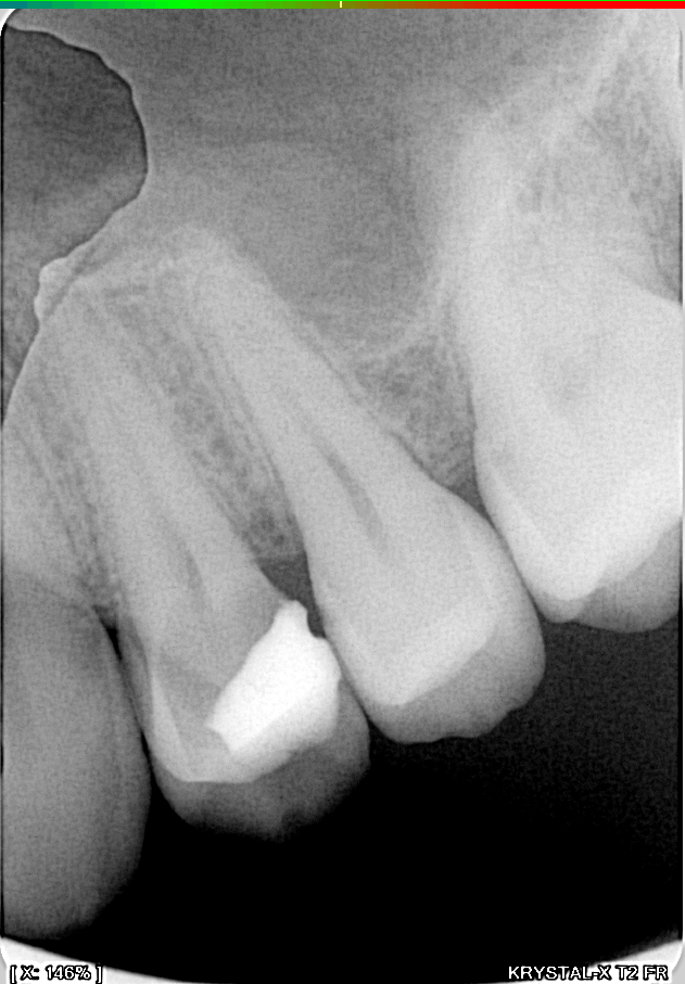
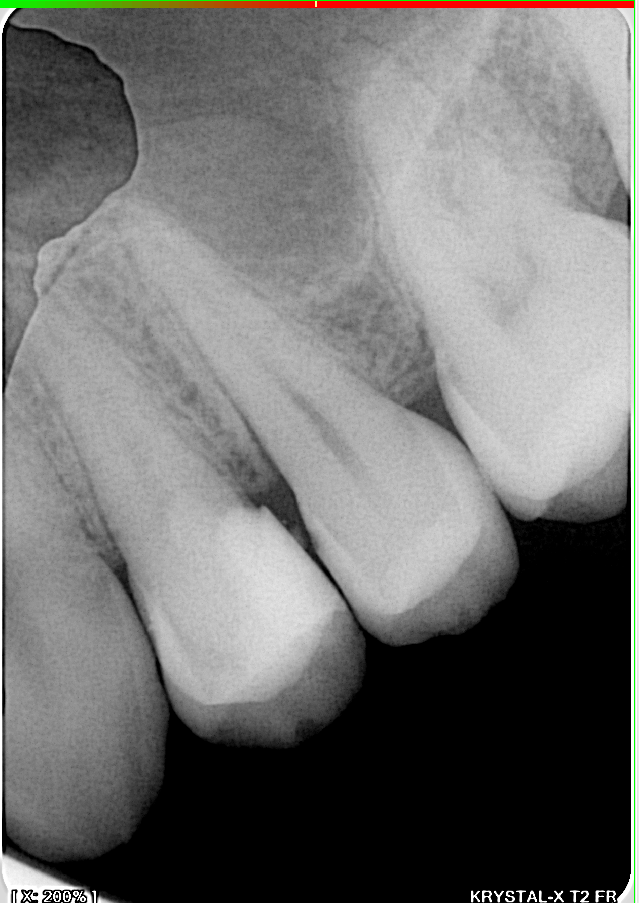
Preoperative radiograph: a significant decay is visible under the composite of the 1st premolar.
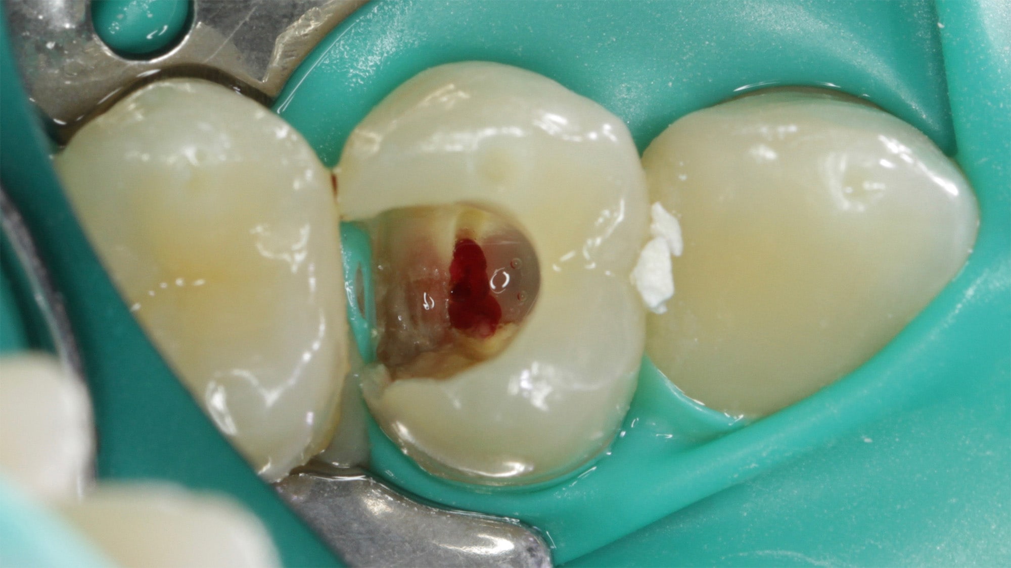
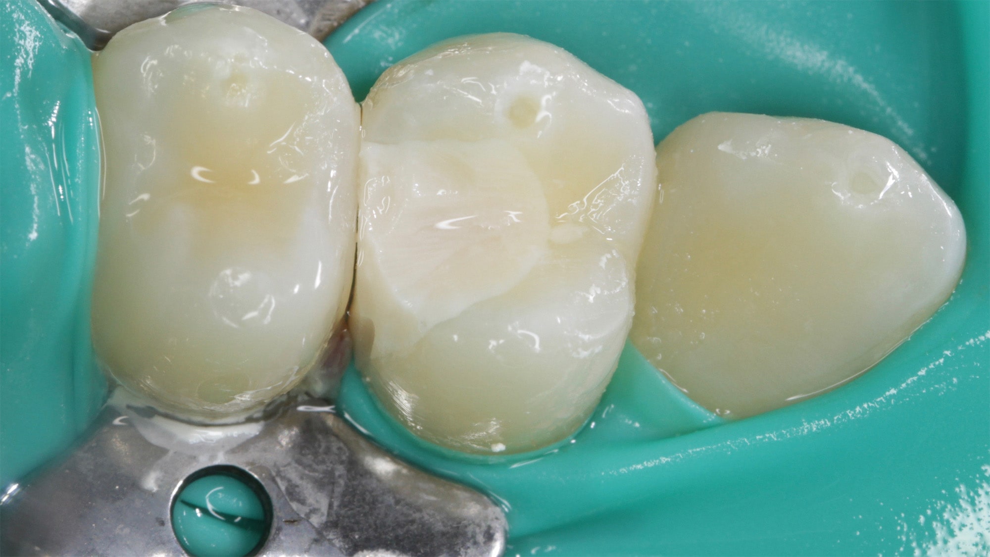
The decay has reached the pulp chamber: the exposed pulp is visible in the center of the tooth, which normally requires complete devitalization of the tooth.
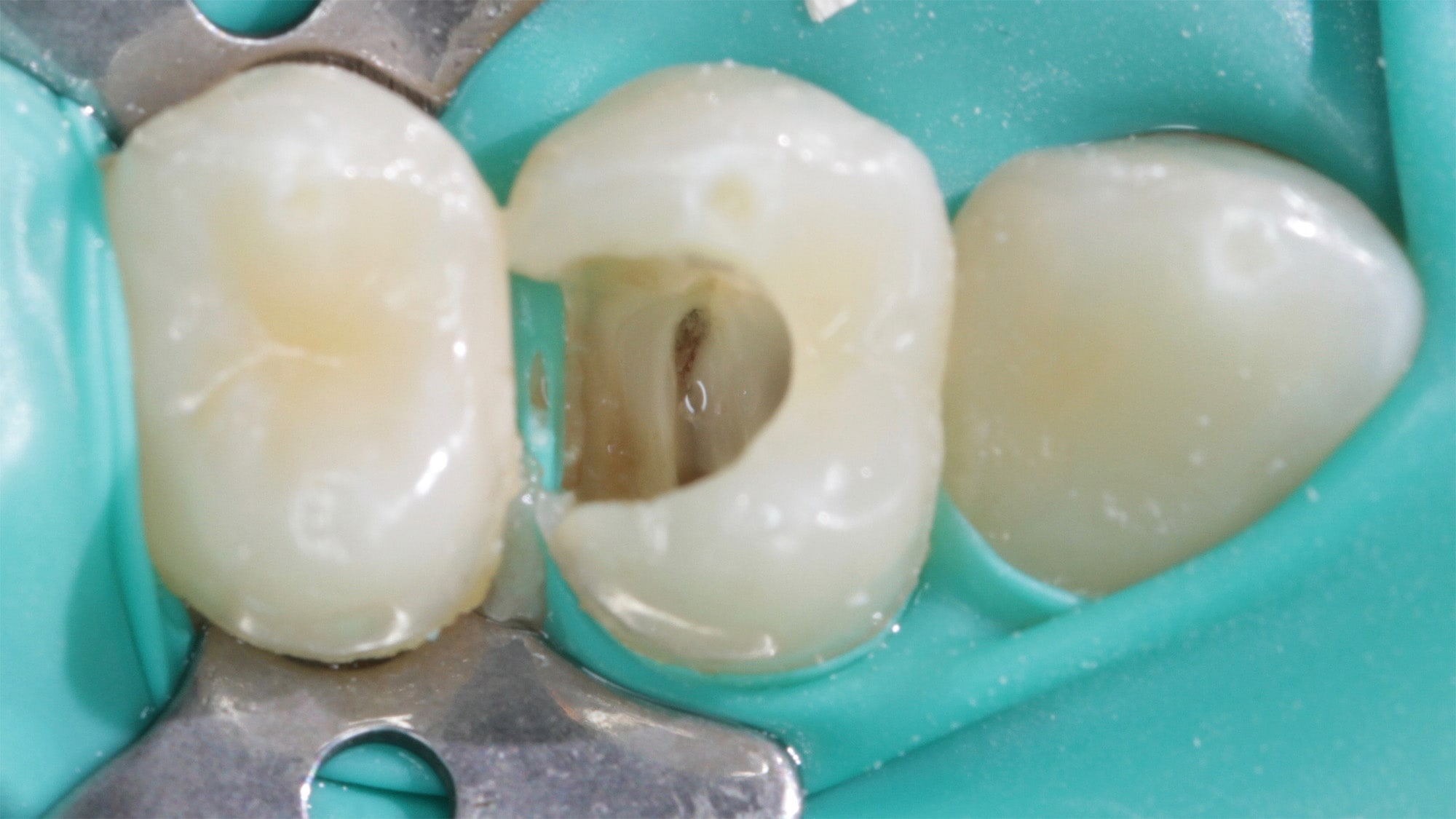
Only the upper part of the pulp is removed and the remaining vital part is kept inside the channels (pulpotomy).
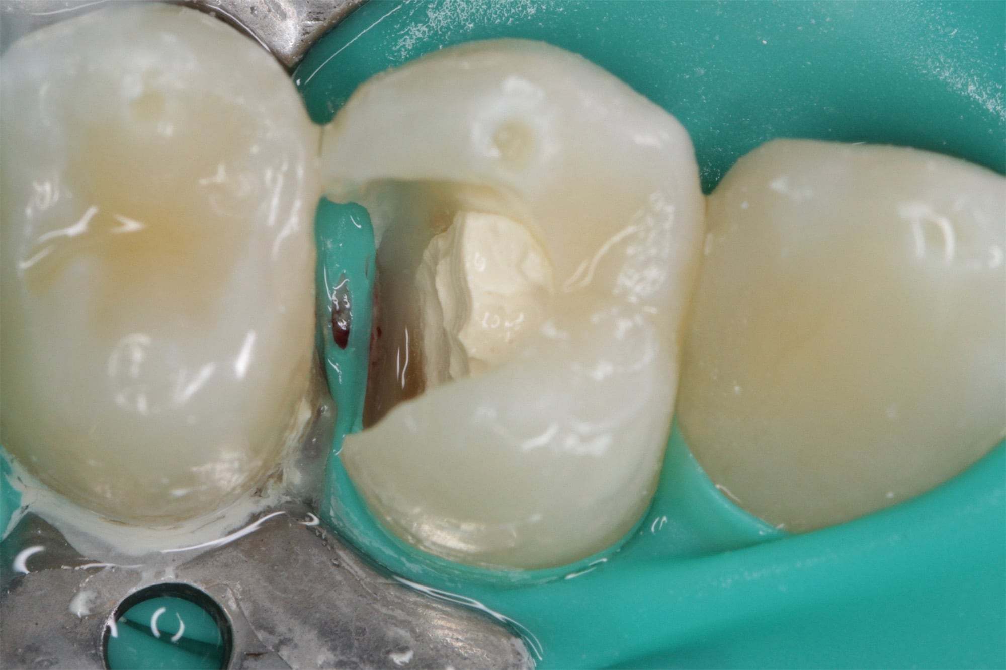
The pulp chamber is closed with biodentine, a highly watertight and biocompatible material to protect the preserved pulp: a partial devitalization is performed and the treatment is less invasive.
A provisional filling is then carried out while waiting to strengthen the tooth with a ceramic glue restoration.