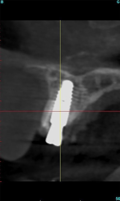
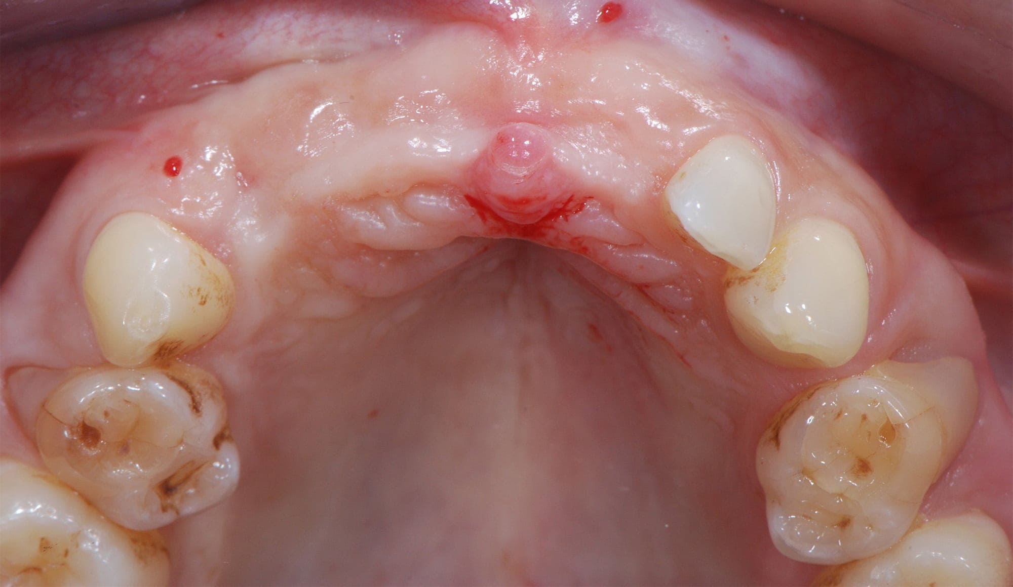
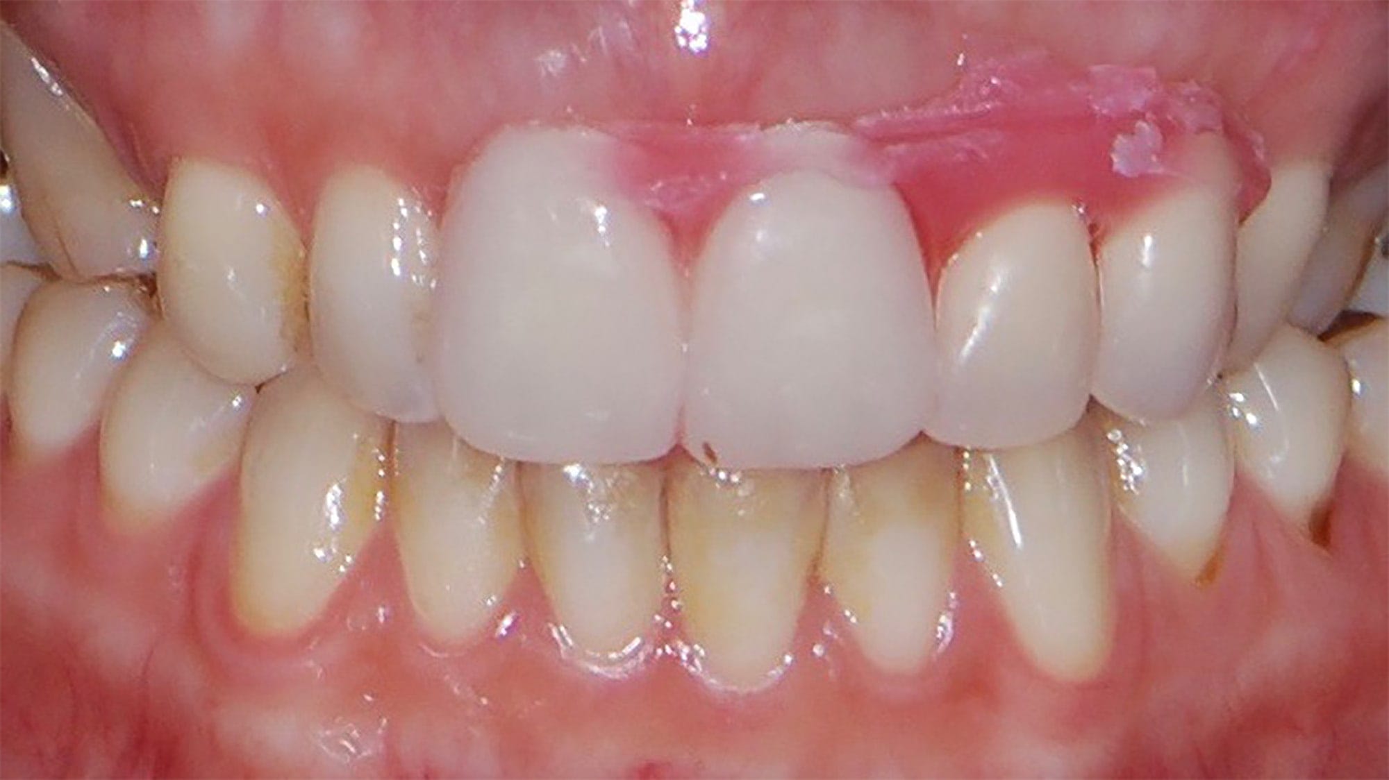
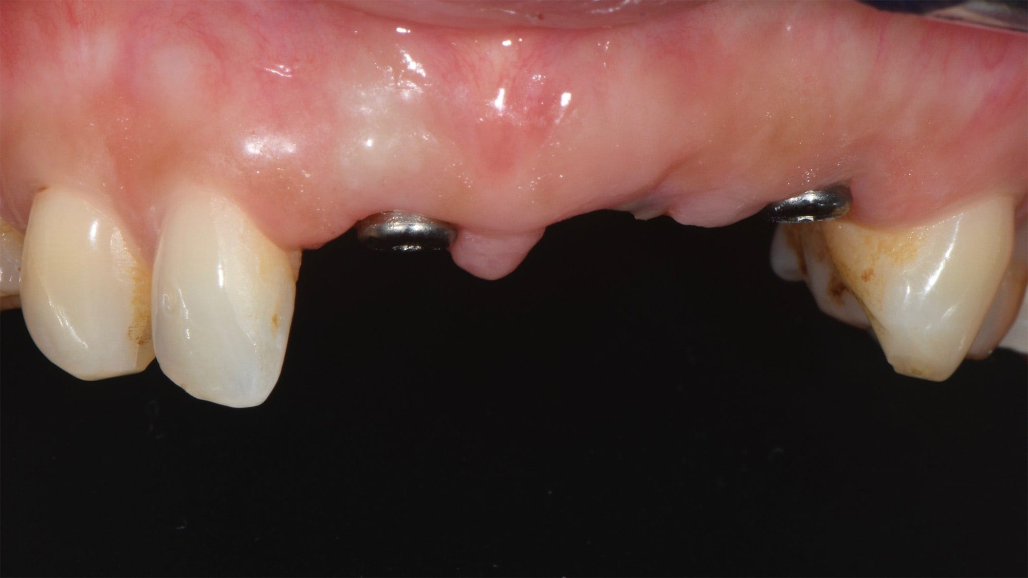
Preoperative setting: Implants are being  considered to replace the absent teeth.

The ideal position of the future teeth is visualized on the patient and on the images of the cone beam thanks to a wax mounting.
Pre-implant planning: the ideal position of the implants is simulated on the images of the beam cone. Future implant crowns appear in white on the images of the beam cone.

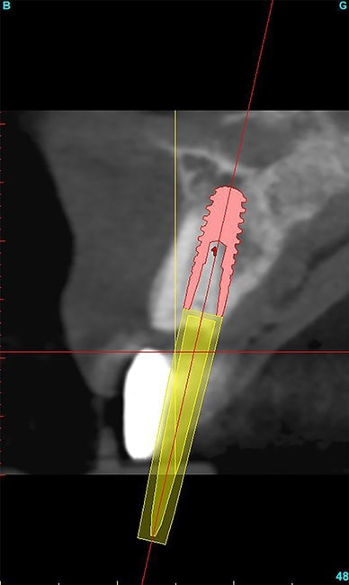
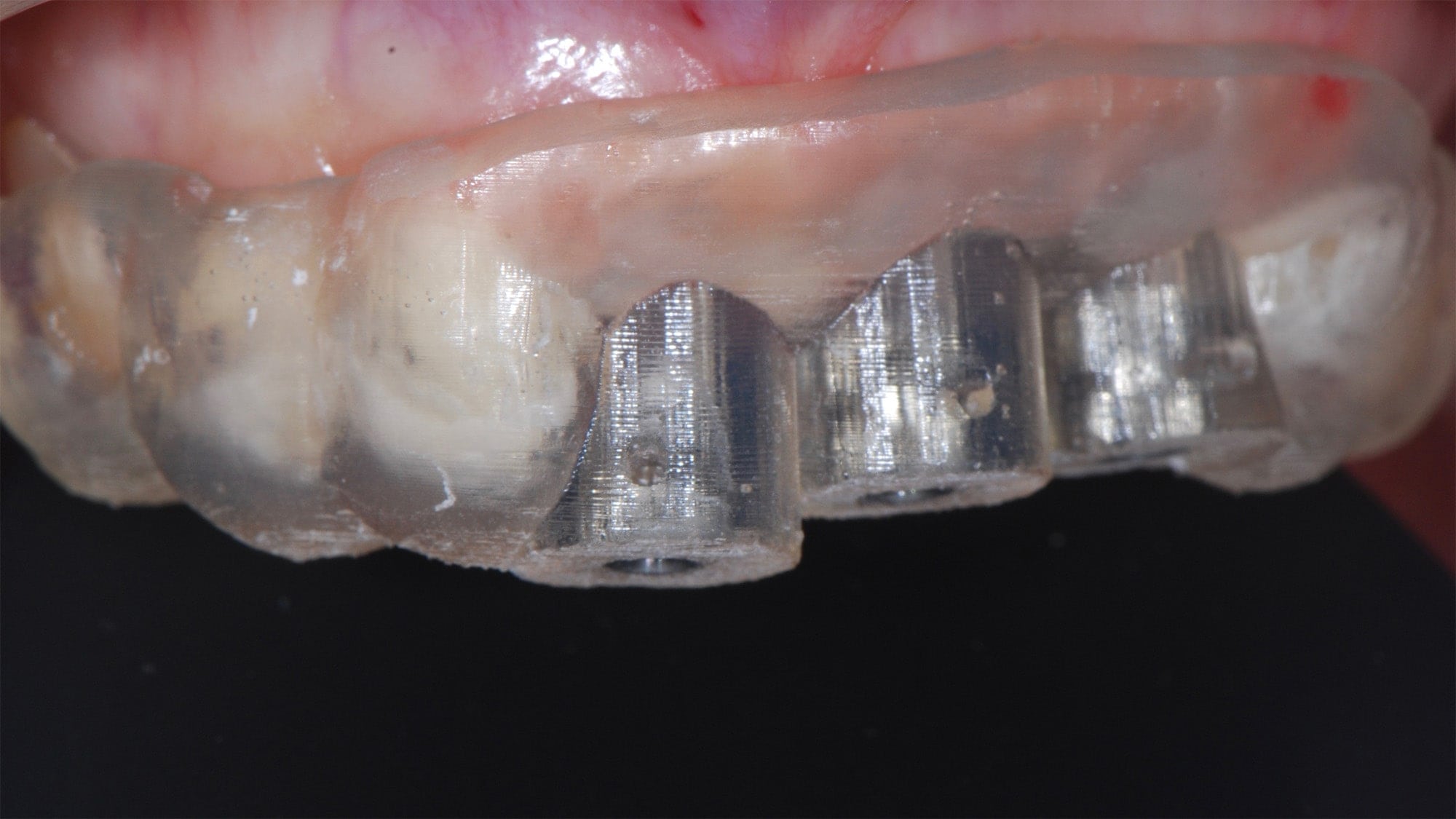
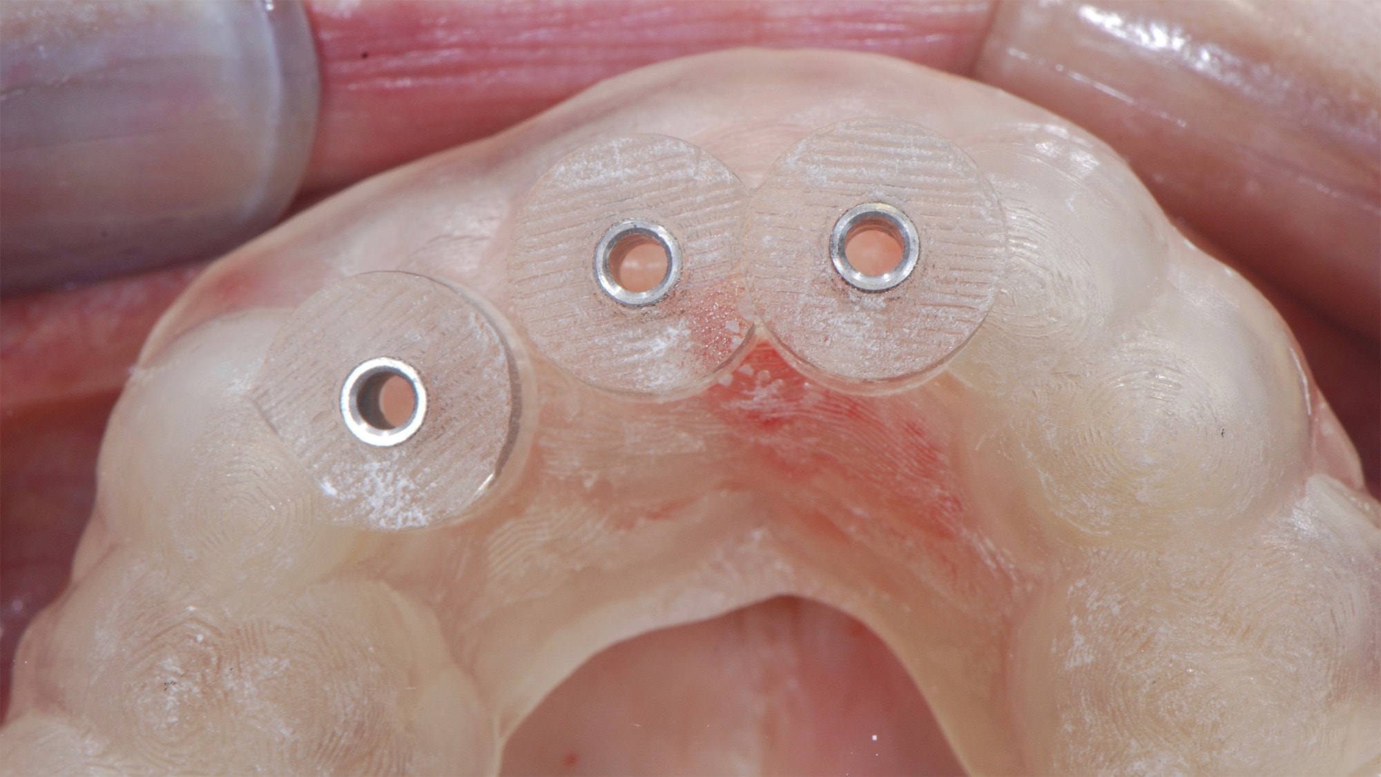
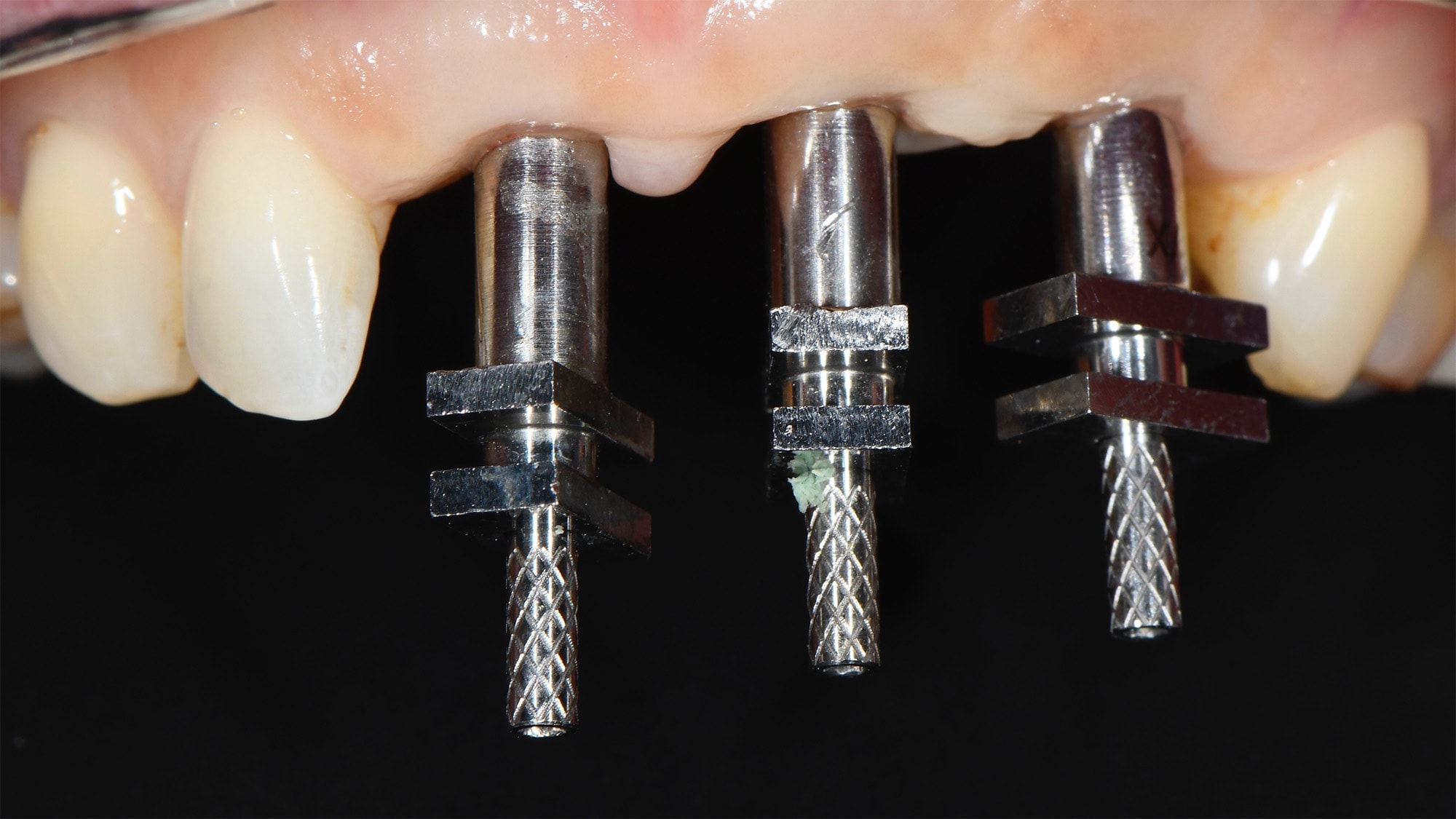
A surgical guide from the pre-implant planning is printed and then tested on the teeth. It allows the surgeon to adequately locate, in the patient’s “oral cavity” , the ideal position for the implants with respect to the bone envelope and the future position of the crowns on implants.

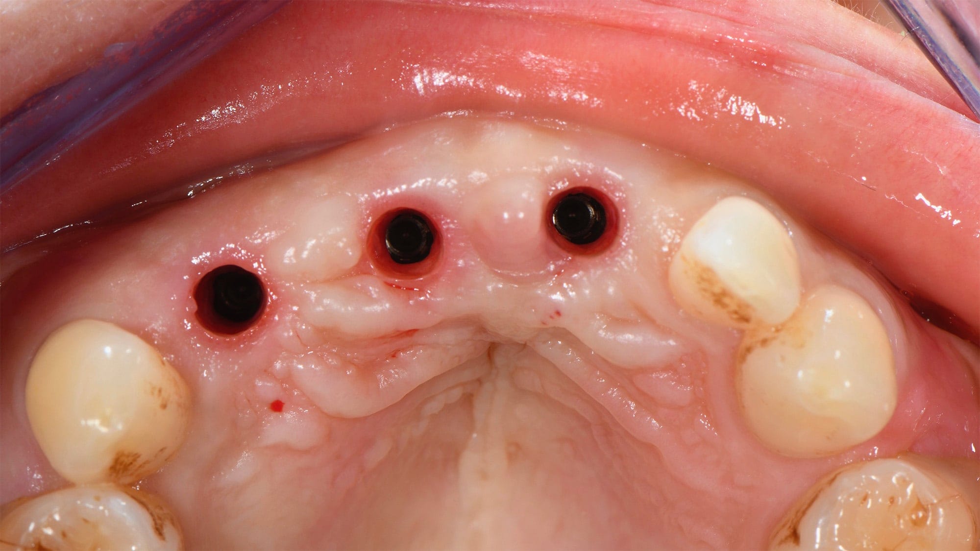
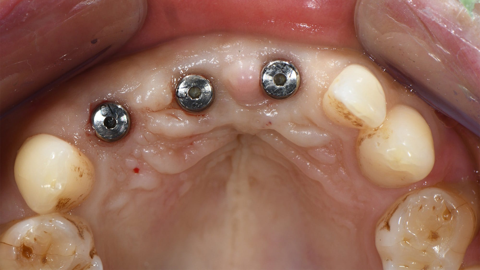
The implants are placed through the guide without “opening” the gum (minimally invasive surgery).

Posted section: the implant is placed exactly as in the image as planned. The residual root has been retained to maintain the bone and gum around the implant crown.
3 months after implant placement, an impression is made to prepare the ceramic crowns on implants (transvissé bridge).
Implanted ceramic bridge on implants (laminated ceramic on zirconia frame).