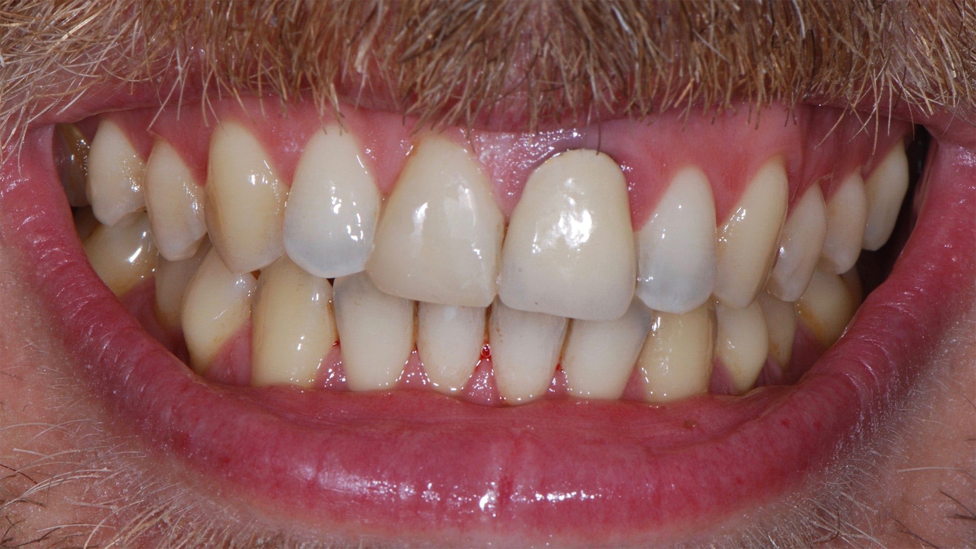
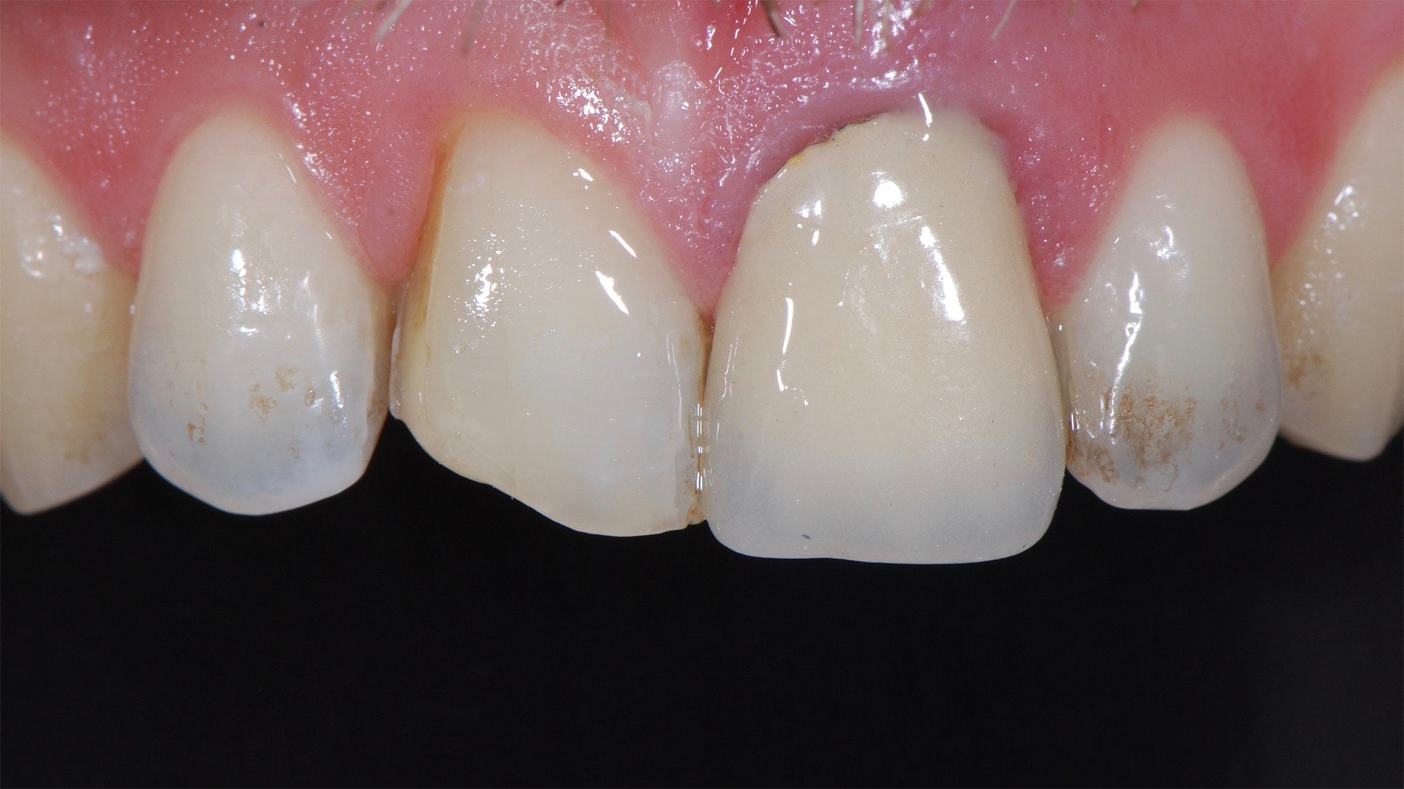
Initial setting: The left central incisor should be extracted because of a significant root infection that has caused significant bone loss around this tooth.
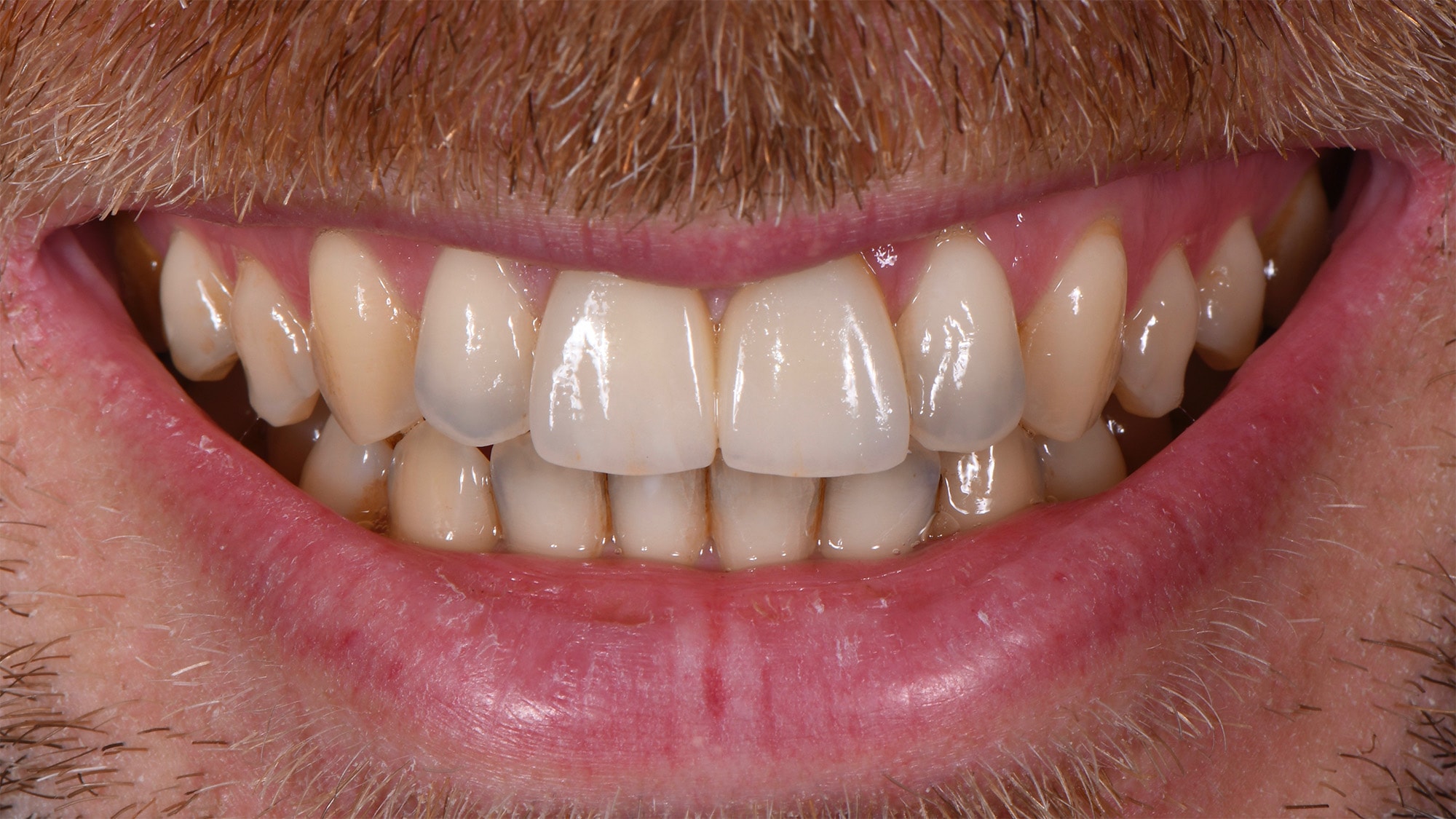
Intra-oral view.
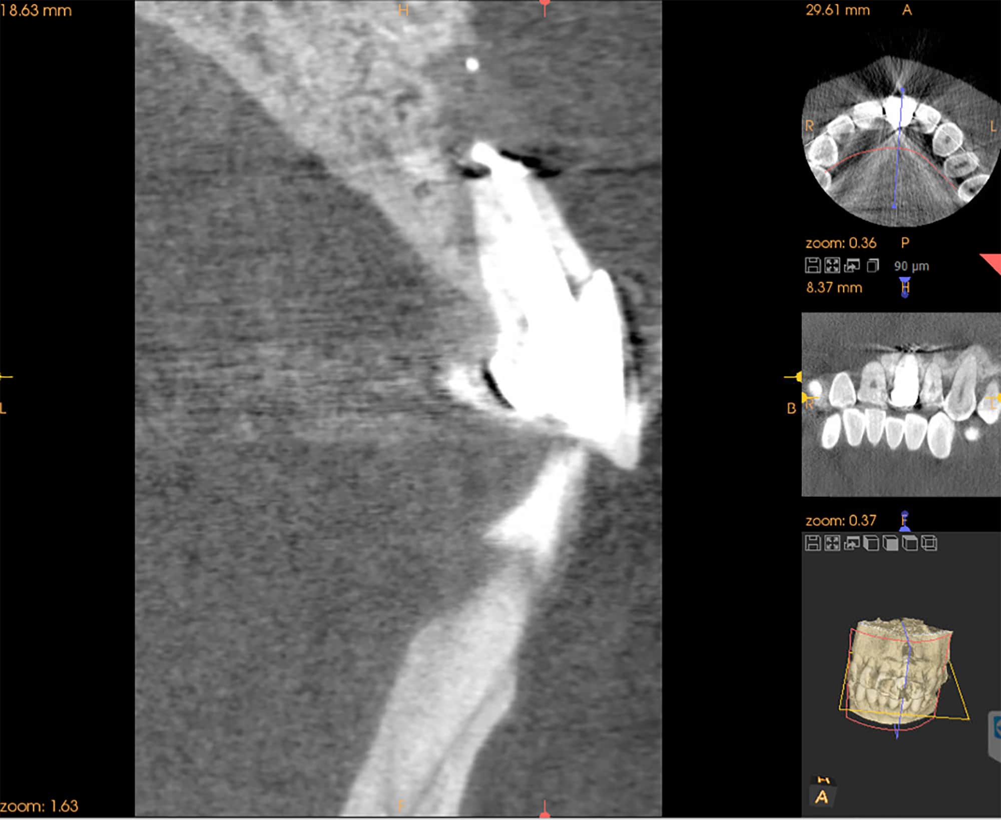
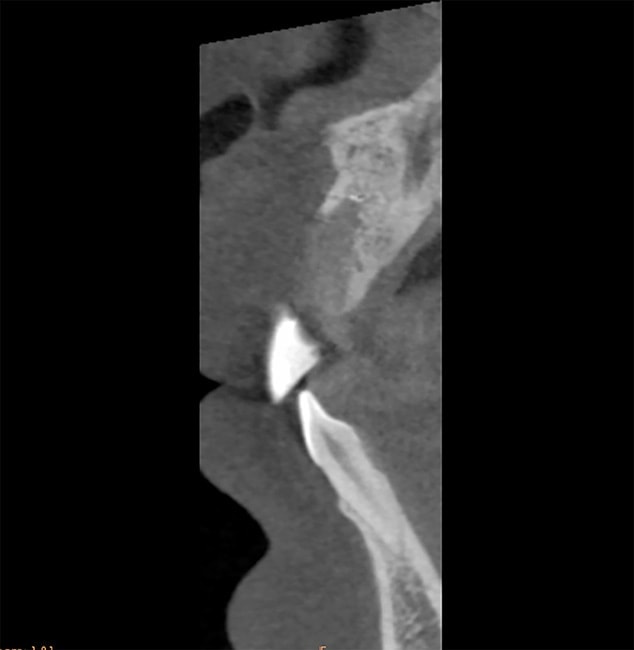
This pre-operative section of the cone beam shows the bone loss around the root of this incisor: this kind of bone defect requires treatment in 3 phases: extraction of the tooth, bone graft 2 months later, implant placement 4 months later, and finally, crown placement on the implant 3 months later. During the 9 months of treatment, the patient will wear a provisional implant attached to the right central incisor.
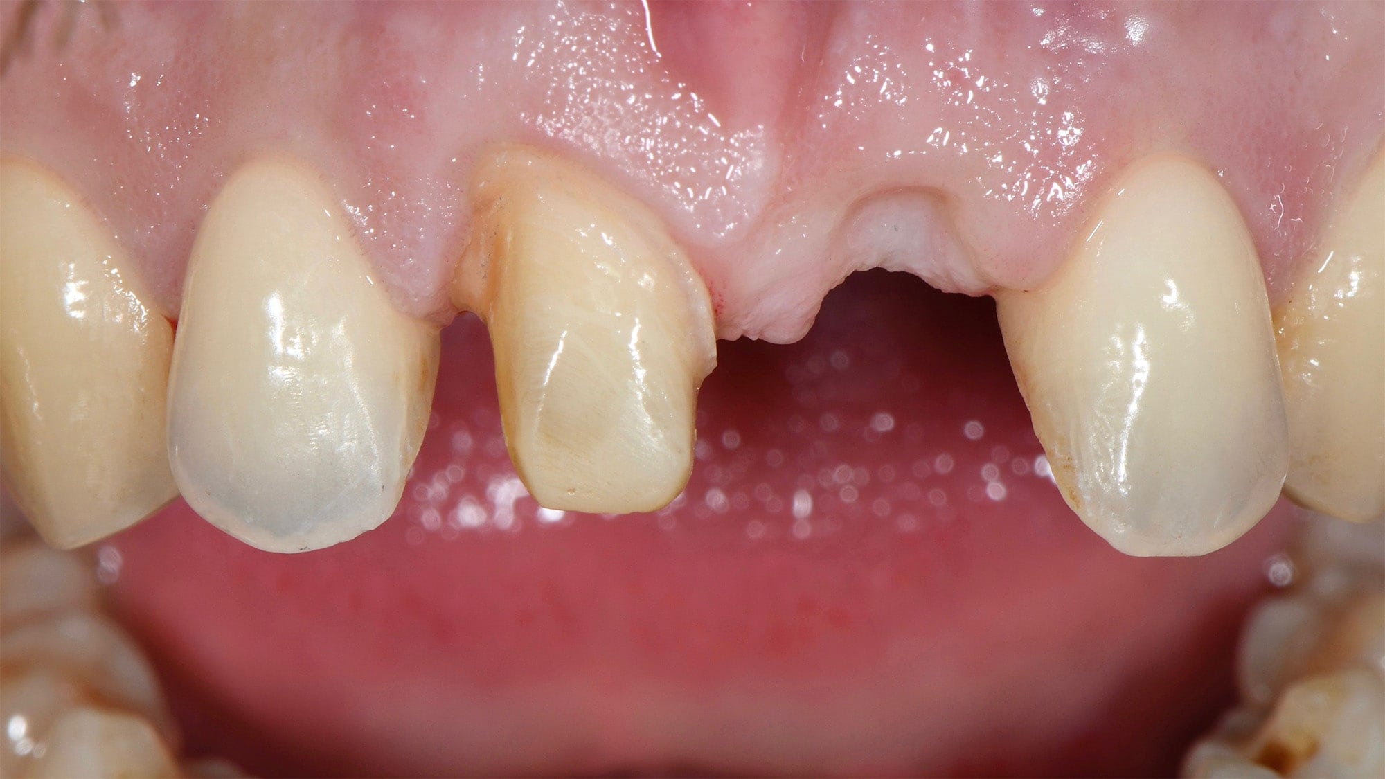
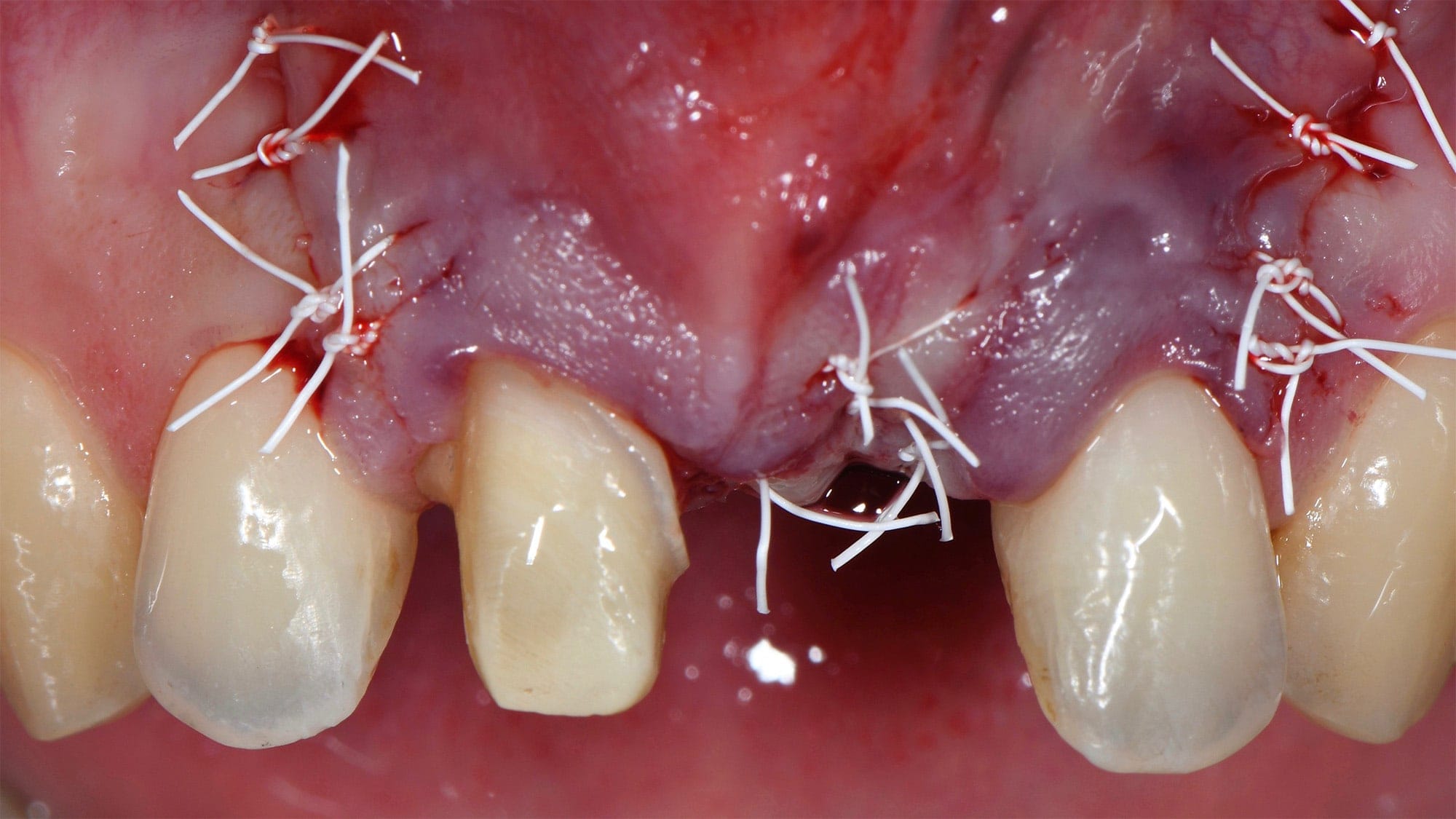
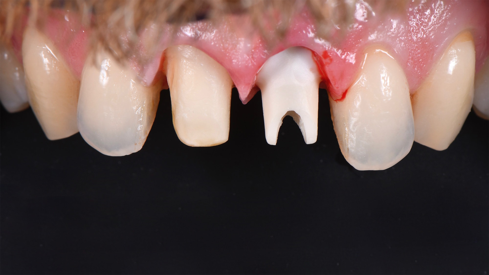
Healing process as seen 2 months after extraction. The right central incisor was prepared to support the fixed provisional implant.
Cone beam slice before bone graft.
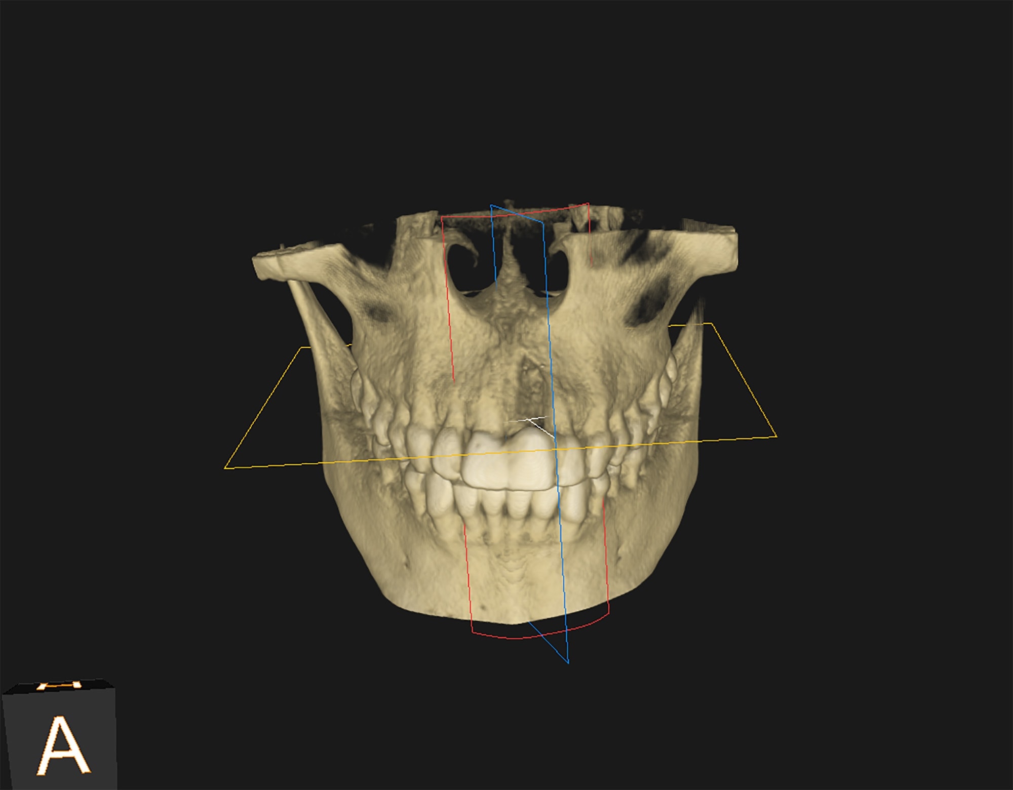
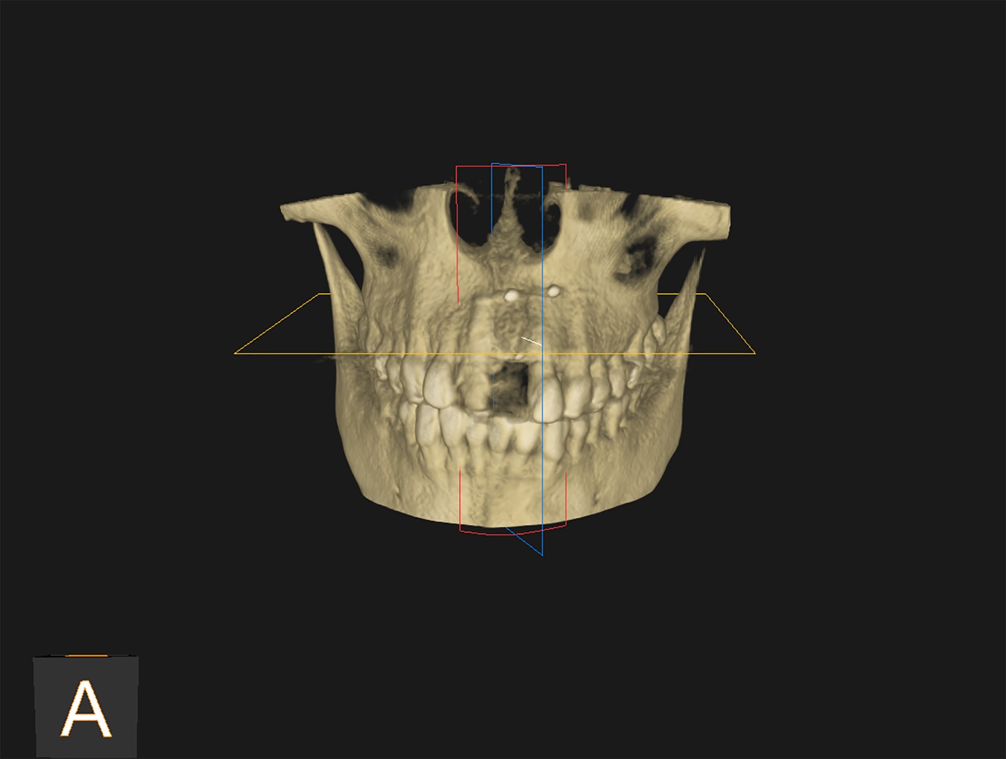
3D view before bone graft: the bone crater is clearly visible at the level of the extracted incisor, which contrasts with the placement of the implant at the time.
Completion of the pre-implant guided bone regeneration.
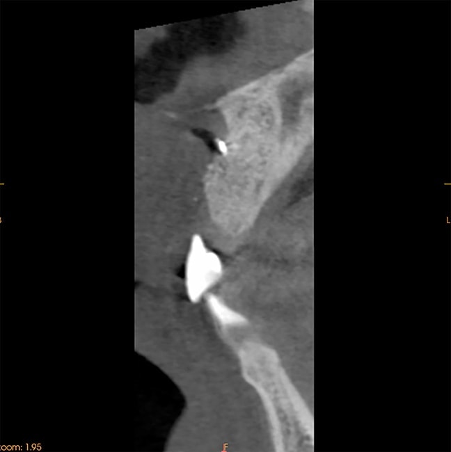
Cone beam slice after bone graft (guided bone regeneration).
3D view after bone graft: a 4-month recovery is required before placing the implant.
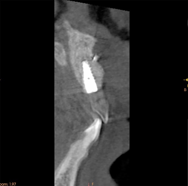
cone beam slice after implant placement.
After bone integration of the implant, a zirconia pillar is transferred to the implant.
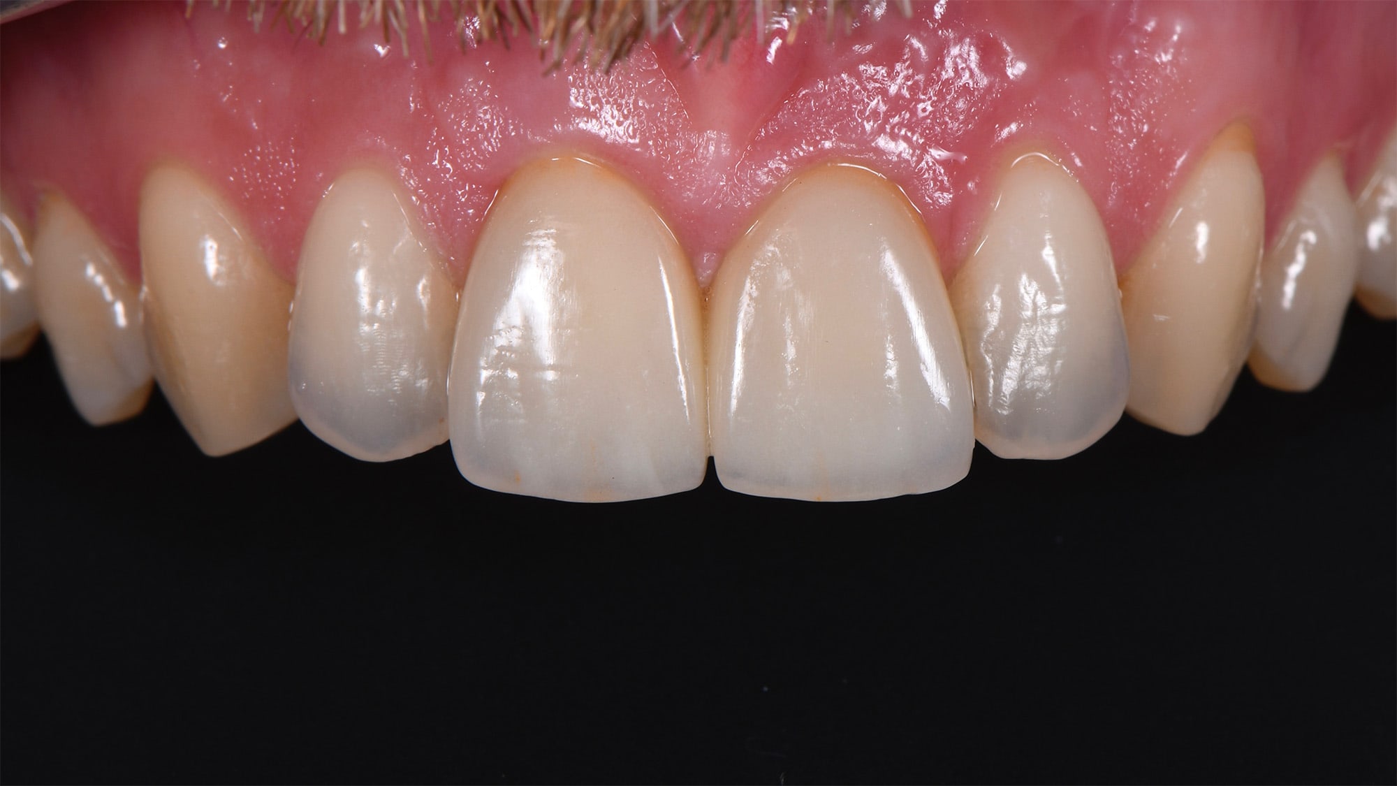
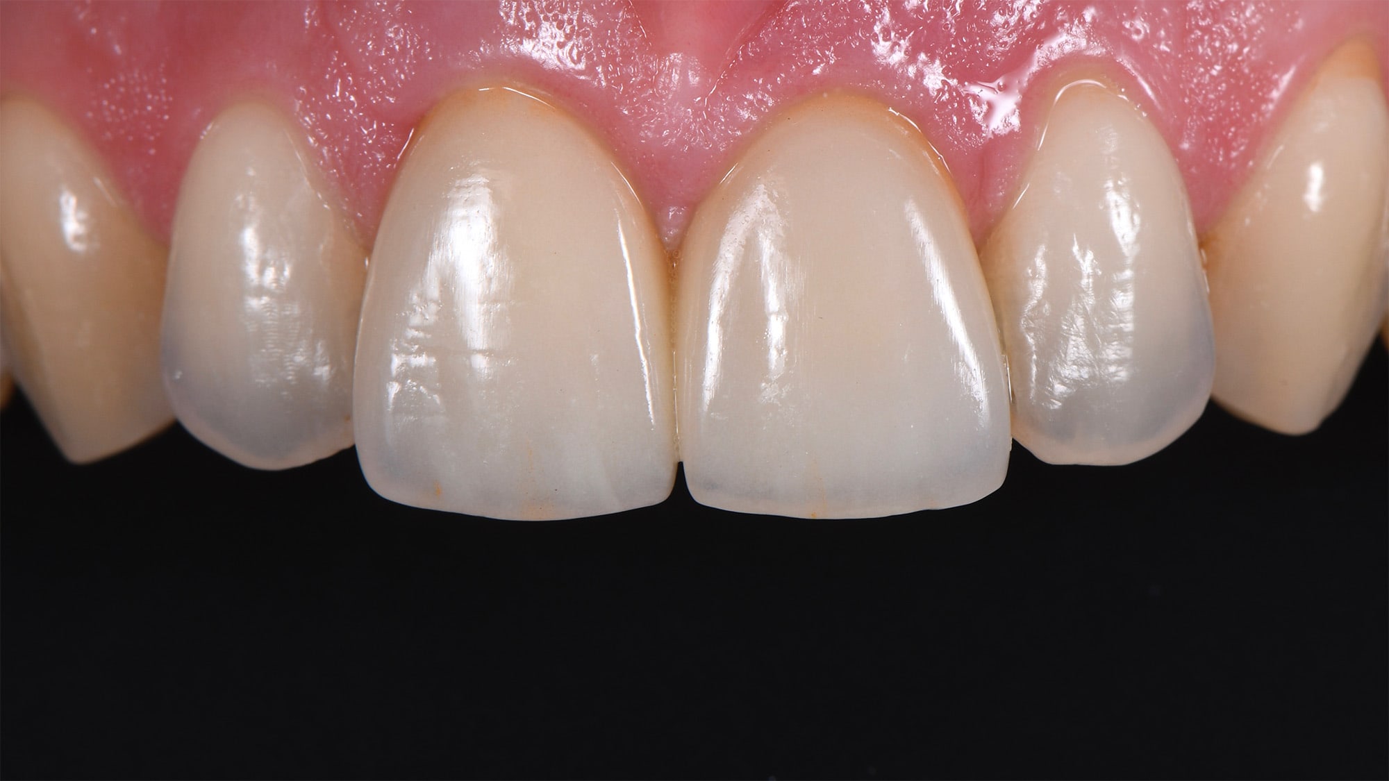
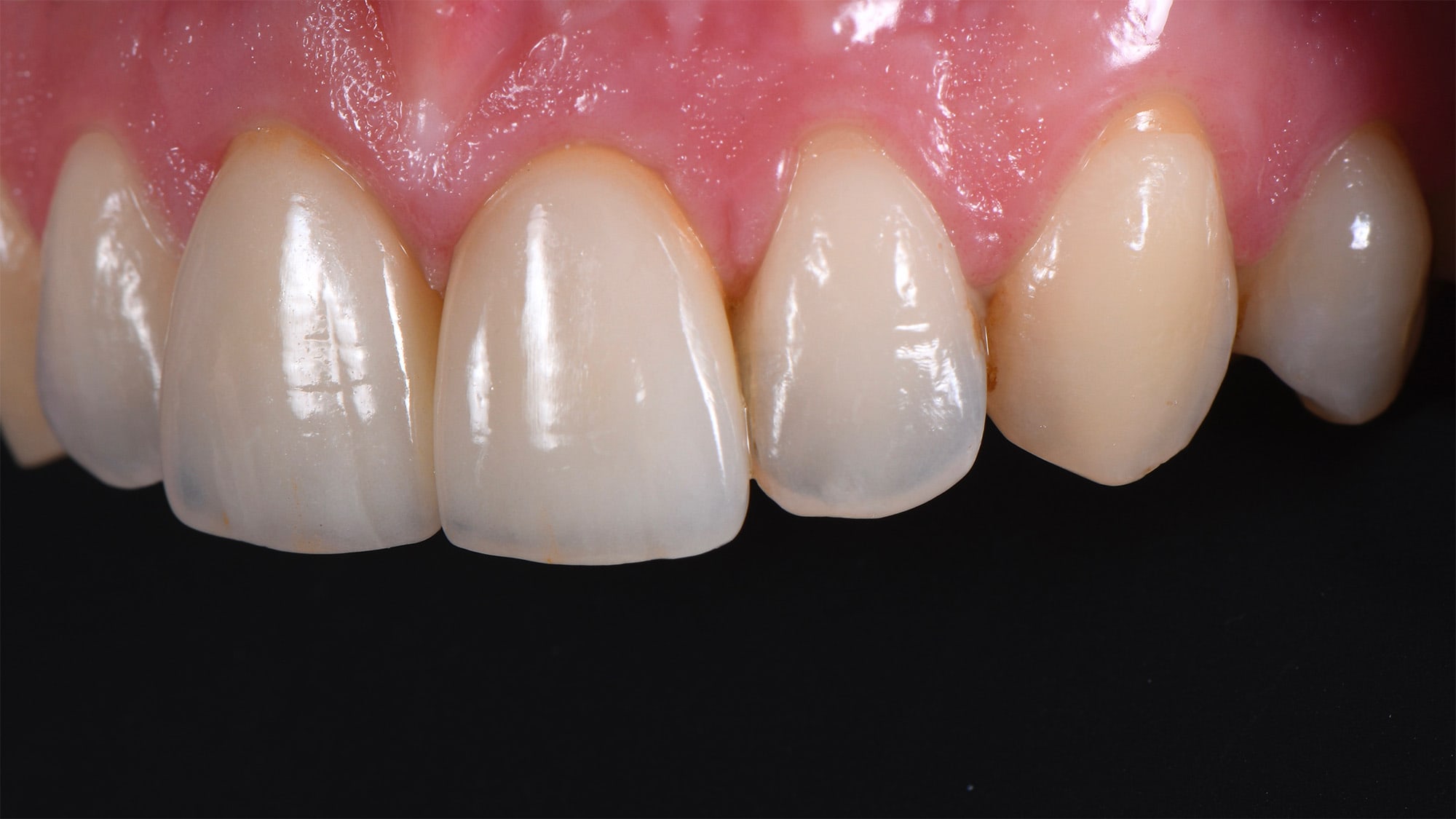
Then the ceramic crowns are laid.
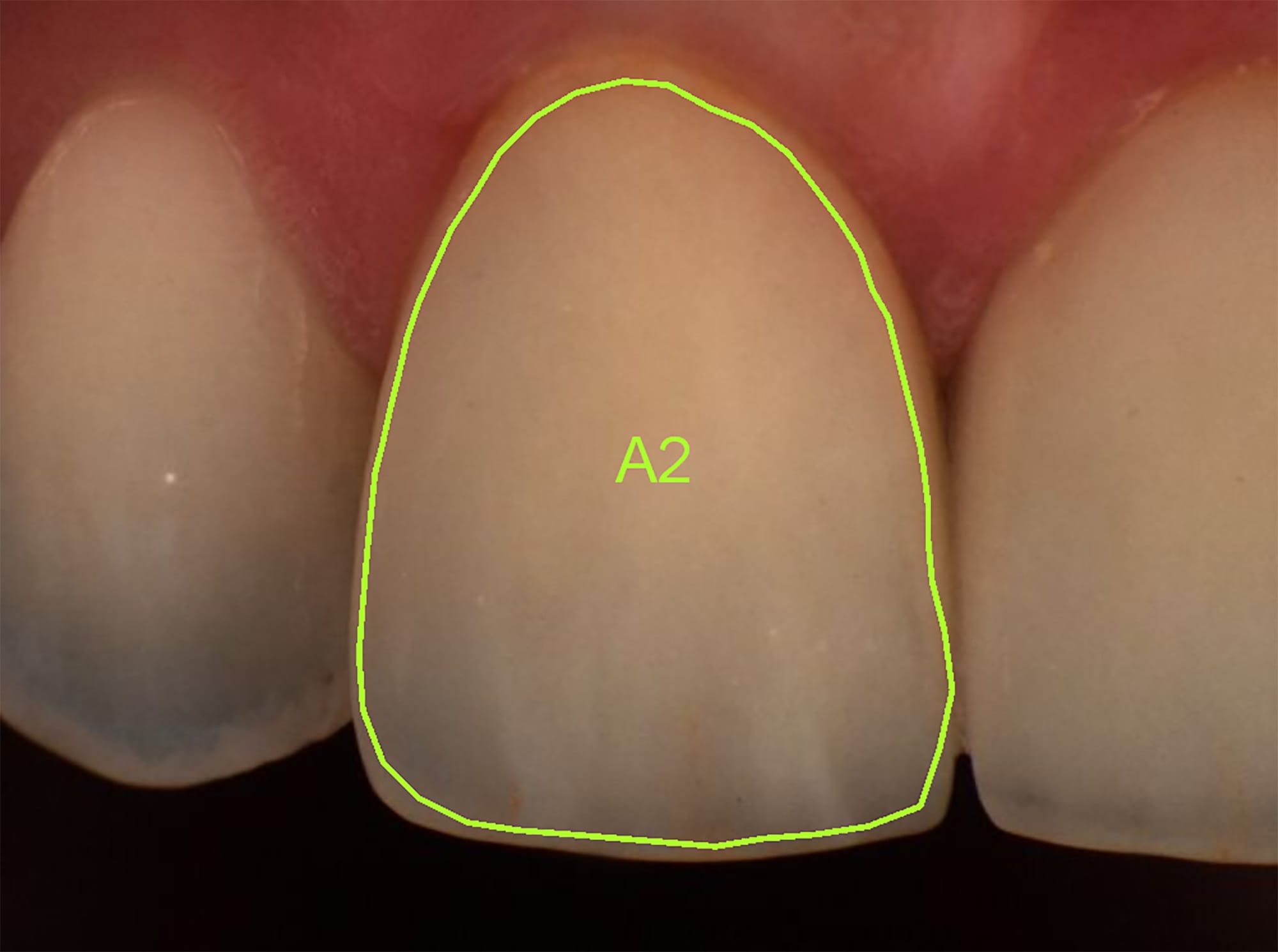
Control of the crown color at the spectrophotometer.