
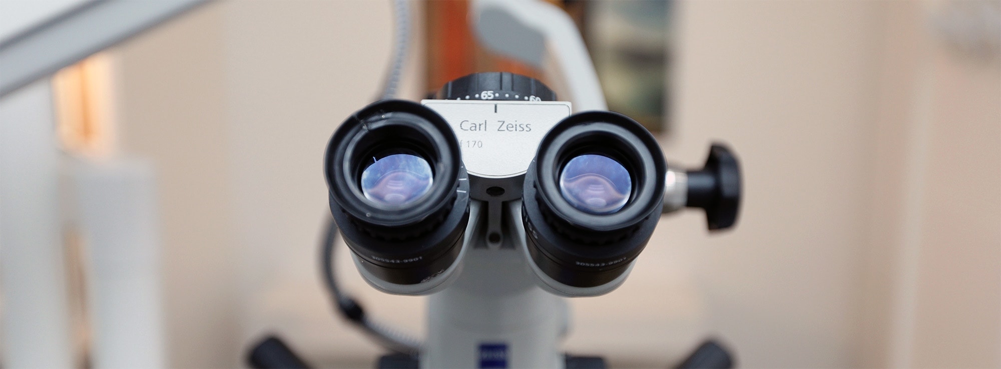
Precision increases the longevity of a treatment, hence the interest of the operating microscope in dentistry.
In endodontics, it allows the practitioner to perform more complex procedures. It should be understood that a channel of a root is 0.5mm in diameter at the entrance and about 80-100um at its apex (the root tip). Thanks to its powerful xenon light and magnification potential of up to x21, the microscope allows the examiner to illuminate the channel along its length, and to see what cannot be seen with the naked eye: fine channels in unusual locations, broken instruments, uncleaned debris, special anatomical variants (late bifurcation of a canal for example) and especially allows one to work with more security. Without a microscope the dentist relies on his touch, he does not see what he does or if he does, in a very, very limited fashion.
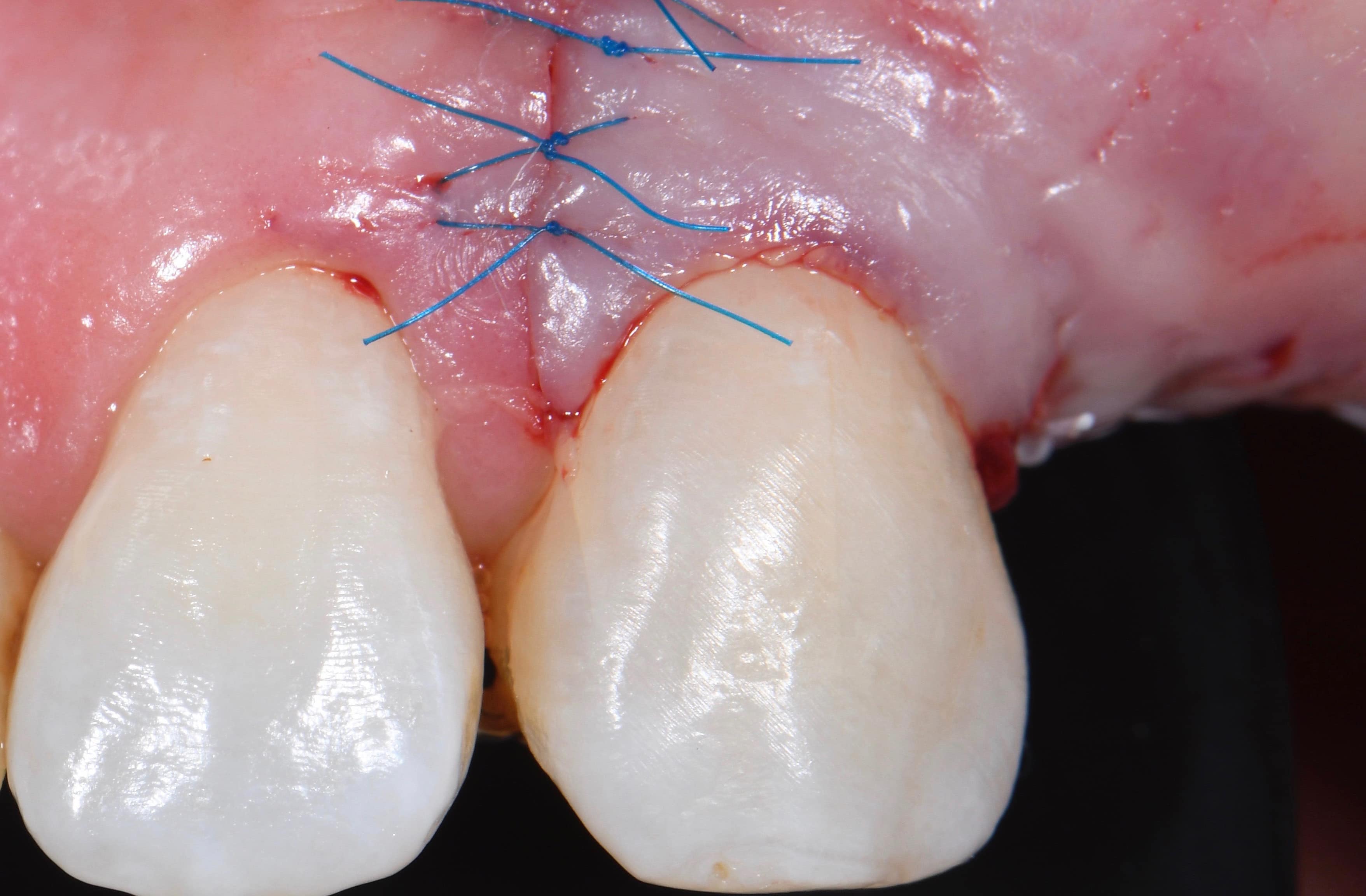
In oral surgery, micro-incisions and micro-sutures accelerate healing: the trauma associated with the incisions is smaller because the micro-blades are very thin. At the time of closure of the site, it is possible to make more stitches because of their extremely thin diameter, which promotes the co-aptation of the edges of the wound and makes the scars less visible.
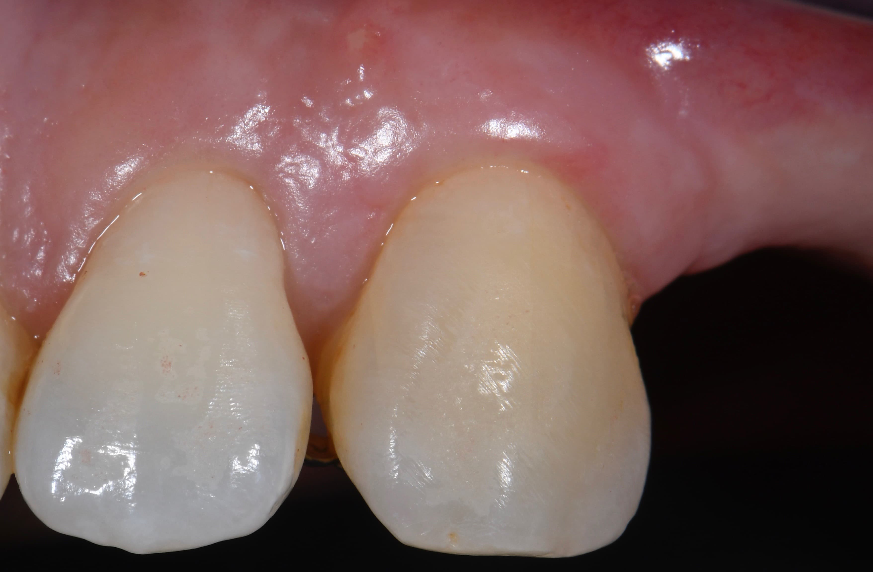
Finally the microscope is also very useful in prosthetics, for the precise completion of minimally invasive dental preparations for ceramic veneers, which optimizes their adaptation and makes the junction between the tooth and the veneer invisible.